WASHINGTON — A nasty stomach virus that can linger on fruits and veggies may have met its match in cold plasma.
In experiments, the ionized gas, created by filtering room-temperature air through an electric field, virtually eliminated norovirus from lettuce, researchers reported February 7 at the American Society for Microbiology Biothreats meeting.
Norovirus is the leading cause of foodborne illness in the United States, infecting more than 20 million people every year. Sterilizing food with heat is one way to kill the virus, but that approach doesn’t work for fresh produce. Cold plasma could be a way to sterilize fruits and vegetables without damaging them, said Hamada Aboubakr, a food microbiologist at the University of Minnesota in St. Paul. Aboubakr and colleagues used a cold plasma device to blast contaminated romaine lettuce leaves and stainless steel surfaces. After five minutes, the plasma wiped out about 99 percent of norovirus particles.
The researchers are testing the device on other foodborne viruses such as hepatitis A, which sickened more than 140 people last year after they ate contaminated strawberries. Unpublished experiments have shown that cold plasma also can destroy drug-resistant bacteria on chicken breasts and leafy greens. Aboubakr hopes to adapt the technology for use in restaurants, on cruise ships and in the produce aisles of grocery stores.
BOSTON — For a lawn that helps the environment — and doesn’t need to be mowed — look to the ocean. Meadows of underwater seagrass plants might lower levels of harmful bacteria in nearby ocean waters, researchers reported February 16 during a news conference at the annual meeting of the American Association for the Advancement of Science. That could make the whole ecosystem — from corals to fish to humans — healthier.
Not truly a grass, seagrasses are flowering plants with long, narrow leaves. They grow in shallow ocean water, spreading into vast underwater lawns. Seagrasses are “a marine powerhouse, almost equal to the rainforest. They’re one of the largest stores of carbon in the ocean,” says study coauthor Joleah Lamb, an ecologist at Cornell University. “But they don’t get a lot of attention.” It’s no secret that seagrasses improve water quality, says James Fourqurean, a biologist at Florida International University in Miami who wasn’t involved in the research, which appears in the Feb. 17 Science. The plants are great at removing excess nitrogen and phosphorus from coastal waters. But now, it seems, they might take away harmful bacteria, too.
A few years ago, Lamb’s colleagues became ill with amoebic dysentery while studying coral reefs in Indonesia, an archipelagic nation that straddles the Indian and Pacific oceans. When a city or village on one of the country’s thousands of islands dumps raw sewage into the ocean, shoreline bacteria populations can spike to dangerous levels. Water sampled close to the shores of four small and densely populated Indonesian islands had 10 times the U.S. Environmental Protection Agency’s recommended exposure limit of Enterococcusbacteria, which can cause illness in humans and often signals the presence of other pathogens. But water collected from offshore tidal flats and coral reefs with seagrass beds had lower levels of the bacteria compared with similar sites without the plants less than 20 meters away. The water had lower levels of numerous bacterial species that can make fish and marine invertebrates sick, too. And field surveys of more than 8,000 coral heads showed that those growing adjacent to or within seagrass beds had fewer diseases than those growing farther away. It’s unclear how far from seagrass beds this cleaner water extends, but the benefits can ripple through the entire ecosystem, Lamb said at the news conference. Healthier corals help protect the islands from erosion. And fish less contaminated with bacteria make a better source of food for people.
Lamb is planning follow-up studies to figure out exactly how the seagrasses clean the water. Like a shag carpet, seagrasses trap small particulates drifting through the ocean and prevent them from flowing on. The plants might ensnare bacteria in the same way, building up biofilms on their blades. Or, she suggests, the leaves could be giving off antimicrobial compounds that directly kill the bacteria.
The findings are one more reason to conserve seagrasses, study coauthor Jeroen van de Water, an ecologist at the Scientific Center of Monaco, said at the news conference. Worldwide, seagrass beds are declining by 7 percent each year, thanks to pollution and habitat loss. And while restoration efforts are underway in some areas, “it’s better to stop what we’re doing to the meadows than to try to replant them,” Lamb added. “Seagrasses are quite particular in the depth they want to be at and the environment they want to have. It’s hard to start doing restoration projects if the environment isn’t exactly what the seagrass prefers.”
Volcanoes that belch hydrogen could bump up the number of potentially habitable planets in the universe.
Ramses Ramirez and Lisa Kaltenegger, both of Cornell University, modeled the atmospheres of planets blemished with hydrogen-spewing volcanoes. These gaseous eruptions could warm planets and ultimately widen a star’s habitable zone, the region where liquid water can exist on a planet’s surface, by about 30 to 60 percent, researchers report in the March 1 Astrophysical Journal Letters. That would be like extending the outer edge of the sun’s habitable zone from about 254 million kilometers — just beyond Mars’ orbit — to 359 million kilometers, or roughly to the asteroid belt between Mars and Jupiter.
Exoplanets that astronomers had previously thought too cold to support life might, in fact, be ripe for habitability if they have hydrogen volcanoes, the researchers say. One example is TRAPPIST-1h, the farthest-out planet identified in an odd system of seven Earth-sized planets 39 light-years from Earth (SN Online: 2/22/17). That world is thought to be icy like Jupiter’s moon Europa.
Adding planets to a star’s habitable zone means more exotic worlds could be targets in the search for signatures of life beyond our solar system. Astronomers plan to search for these signatures with the James Webb Space Telescope, slated to launch in 2018, and later with the European Extremely Large Telescope, scheduled to begin operations in 2024.
Just six weeks of training can turn average people into memory masters.
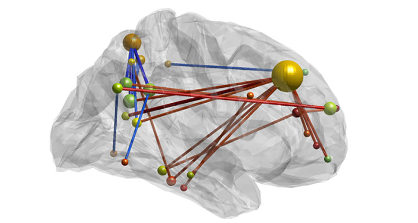
Boosting these prodigious mnemonic skills came with overhauls in brain activity, resulting in brains that behaved more like those of experts who win World Memory Championships competitions, scientists report March 8 in Neuron.
The findings are notable because they show just how remarkably adaptable the human brain is, says neuroscientist Craig Stark of the University of California, Irvine. “The brain is plastic,” he says. “Through use, it changes.” It’s not yet clear how long the changes in the newly trained brains last, but the memory gains persisted for four months.
In an initial matchup, a group of 17 memory experts, people who place high in World Memory Championships, throttled a group of people with average memories. Twenty minutes after seeing a list of 72 words, the experts remembered an average of 70.8 words; the nonexperts caught, on average, only 39.9 words.
In subsequent matchups, some nonexperts got varying levels of help. Fifty-one novices were split into three groups. A third of these people spent six weeks learning the method of loci, a memorization strategy used by ancient Greek and Roman orators. To use the technique, a person must imagine an elaborate mental scene, such as a palace or a familiar walking path, and populate it with memorable items. New information can then be placed onto this scaffold, offering a way to quickly “see” long lists of items.
Other participants spent six weeks training to improve short-term memory, performing a tricky task that required people to simultaneously keep track of series of locations they see and numbers they hear. The rest of the participants had no training at all.
After the training, the people who learned the method of loci performed nearly as well as the memory experts. But the rest didn’t show such improvement. Study coauthor Martin Dresler, a neuroscientist at the Radboud University Medical Center in the Netherlands, knew that the method of loci works quite well; he wasn’t surprised to see those memory scores spike. To him, the more interesting changes happened in the trained people’s brains. Before and after training, nonexperts underwent scans that pinpointed brain areas that were active at the same time, an indication that these brain areas work together closely. Dresler and colleagues looked at 2,485 connections in brain networks important for memory and visual and spatial thinking. Training in the method of loci seemed to reconfigure many of those connections, making some of the connections stronger and others weaker. The overall effect of training was to make brains “look like those of the world’s best memorizers,” Dresler says. The results suggest that large-scale changes across the brain, as opposed to changes in individual areas, drive the increased memory capacity.
These new memory skills were still obvious four months after training ended, particularly for the people whose brain behavior became more similar to that of the memory experts. The researchers didn’t scan participants’ brains four months out, so they don’t know whether the brain retains its reshaped connections. No such brain changes or big increases in memory skills were seen in the other groups.
Memorization techniques have been criticized as interesting tricks that have little use in real life. But “that’s not the case,” Dresler says. Boris Konrad, a coauthor of the study also at Radboud, is a memory master who trained in the method of loci. The technique “really helped him get much better grades” in physics and other complex studies, Dresler says.
Improvements in mnemonic memory, like other types of cognitive training, might not improve a broader range of thinking skills. The current study can’t answer bigger questions about whether brain training has more general benefits.
Not too long ago, the internet was stationary. Most often, we’d browse the Web from a desktop computer in our living room or office. If we were feeling really adventurous, maybe we’d cart our laptop to a coffee shop. Looking back, those days seem quaint.
Today, the internet moves through our lives with us. We hunt Pokémon as we shuffle down the sidewalk. We text at red lights. We tweet from the bathroom. We sleep with a smartphone within arm’s reach, using the device as both lullaby and alarm clock. Sometimes we put our phones down while we eat, but usually faceup, just in case something important happens. Our iPhones, Androids and other smartphones have led us to effortlessly adjust our behavior. Portable technology has overhauled our driving habits, our dating styles and even our posture. Despite the occasional headlines claiming that digital technology is rotting our brains, not to mention what it’s doing to our children, we’ve welcomed this alluring life partner with open arms and swiping thumbs.
Scientists suspect that these near-constant interactions with digital technology influence our brains. Small studies are turning up hints that our devices may change how we remember, how we navigate and how we create happiness — or not. Somewhat limited, occasionally contradictory findings illustrate how science has struggled to pin down this slippery, fast-moving phenomenon. Laboratory studies hint that technology, and its constant interruptions, may change our thinking strategies. Like our husbands and wives, our devices have become “memory partners,” allowing us to dump information there and forget about it — an off-loading that comes with benefits and drawbacks. Navigational strategies may be shifting in the GPS era, a change that might be reflected in how the brain maps its place in the world. Constant interactions with technology may even raise anxiety in certain settings.
Yet one large study that asked people about their digital lives suggests that moderate use of digital technology has no ill effects on mental well-being.
The question of how technology helps and hinders our thinking is incredibly hard to answer. Both lab and observational studies have drawbacks. The artificial confines of lab experiments lead to very limited sets of observations, insights that may not apply to real life, says experimental psychologist Andrew Przybylski of the University of Oxford. “This is a lot like drawing conclusions about the effects of baseball on players’ brains after observing three swings in the batting cage.”
Observational studies of behavior in the real world, on the other hand, turn up associations, not causes. It’s hard to pull out real effects from within life’s messiness. The goal, some scientists say, is to design studies that bring the rigors of the lab to the complexities of real life, and then to use the resulting insights to guide our behavior. But that’s a big goal, and one that scientists may never reach.
Evolutionary neurobiologist Leah Krubitzer is comfortable with this scientific ambiguity. She doesn’t put a positive or negative value on today’s digital landscape. Neither good nor bad, it just is what it is: the latest iteration on the continuum of changing environments, says Krubitzer, of the University of California, Davis.
“I can tell you for sure that technology is changing our brains,” she says. It’s just that so far, no one knows what those changes mean.
Of course, nearly everything changes the brain. Musical training reshapes parts of the brain. Learning the convoluted streets of London swells a mapmaking structure in the brains of cabbies. Even getting a good night’s sleep changes the brain. Every aspect of our environment can influence brain and behaviors. In some ways, digital technology is no different. Yet some scientists suspect that there might be something particularly pernicious about digital technology’s grip on the brain.
“We are information-seeking creatures,” says neuroscientist Adam Gazzaley of the University of California, San Francisco. “We are driven to it in very powerful ways.” Today’s digital tools give us unprecedented exposure to information that doesn’t wait for you to seek it out; it seeks you out, he says. That pull is nearly irresistible.
Despite the many unanswered questions about whether our digital devices are influencing our brains and behaviors, and whether for good or evil, technology is galloping ahead. “We should have been asking ourselves [these sorts of questions] in the ’70s or ’80s,” Krubitzer says. “It’s too late now. We’re kind of closing the barn doors after the horses got out.” Attention grabber One way in which today’s digital technology is distinct from earlier advances (like landline telephones) is the sheer amount of time people spend with it. In just a decade, smartphones have saturated the market, enabling instant internet access to an estimated 2 billion people around the world. In one small study reported in 2015, 23 adults, ages 18 to 33, spent an average of five hours a day on their phones, broken up into 85 distinct daily sessions. When asked how many times they thought they used their phones, participants underestimated by half.
In a different study, Larry Rosen, a psychologist at California State University, Dominguez Hills, used an app to monitor how often college students unlocked their phones. The students checked their phones an average of 60 times a day, each session lasting about three to four minutes for a total of 220 minutes a day. That’s a lot of interruption, Rosen says. Smartphones are “literally omnipresent 24-7, and as such, it’s almost like an appendage,” he says. And often, we are compelled to look at this new, alluring rectangular limb instead of what’s around us. “This device is really powerful,” Rosen says. “It’s really influencing our behavior. It’s changed the way we see the world.”
Technology does that. Printing presses, electricity, televisions and telephones all shifted people’s habits drastically, Przybylski says. He proposes that the furor over digital technology melting brains and crippling social lives is just the latest incarnation of the age-old fear of change. “You have to ask yourself, ‘Is there something magical about the power of an LCD screen?’ ” Przybylski says.
Yet some researchers suspect that there is something particularly compelling about this advance. “It just feels different. Computers and the internet and the cloud are embedded in our lives,” says psychologist Benjamin Storm of the University of California, Santa Cruz. “The scope of the amount of information we have at our fingertips is beyond anything we’ve ever experienced. The temptation to become really reliant on it seems to be greater.”
Memory outsourcing Our digital reliance may encourage even more reliance, at least for memory, Storm’s work suggests. Sixty college undergraduates were given a mix of trivia questions — some easy, some hard. Half of the students had to answer the questions on their own; the other half were told to use the internet. Later, the students were given an easier set of questions, such as “What is the center of a hurricane called?” This time, the students were told they could use the internet if they wanted.
People who had used the internet initially were more likely to rely on internet help for the second, easy set of questions, Storm and colleagues reported online last July in Memory. “People who had gotten used to using the internet continued to do so, even though they knew the answer,” Storm says. This kind of overreliance may signal a change in how people use their memory. “No longer do we just rely on what we know,” he says. That work builds on results published in a 2011 paper in Science . A series of experiments showed that people who expected to have access to the internet later made less effort to remember things . In this way, the internet has taken the place formerly filled by spouses who remember birthdays, grandparents who remember recipes and coworkers who remember the correct paperwork codes — officially known as “transactive memory partners.” “We are becoming symbiotic with our computer tools,” Betsy Sparrow, then at Columbia University, and colleagues wrote in 2011. “The experience of losing our internet connection becomes more and more like losing a friend. We must remain plugged in to know what Google knows.”
That digital crutch isn’t necessarily a bad thing, Storm points out. Human memory is notoriously squishy, susceptible to false memories and outright forgetting. The internet, though imperfect, can be a resource of good information. And it’s not clear, he says, whether our memories are truly worse, or whether we perform at the same level, but just reach the answer in a different way.
“Some people think memory is absolutely declining as a result of us using technology,” he says. “Others disagree. Based on the current data, though, I don’t think we can really make strong conclusions one way or the other.”
The potential downsides of this memory outsourcing are nebulous, Storm says. It’s possible that digital reliance influences — and perhaps even weakens — other parts of our thinking. “Does it change the way we learn? Does it change the way we start to put information together, to build our own stories, to generate new ideas?” Storm asks. “There could be consequences that we’re not necessarily aware of yet.”
Research by Gazzaley and others has documented effects of interruptions and multitasking, which are hard to avoid with incessant news alerts, status updates and Instagrams waiting in our pockets. Siphoning attention can cause trouble for a long list of thinking skills, including short- and long-term memory, attention, perception and reaction time. Those findings, however, come from experiments in labs that ask a person to toggle between two tasks while undergoing a brain scan, for instance. Similar effects have not been as obvious for people going about their daily lives, Gazzaley says. But he is convinced that constant interruptions — the dings and buzzes, our own restless need to check our phones — are influencing our ability to think.
Making maps Consequences of technology are starting to show up for another cognitive task — navigating, particularly while driving. Instead of checking a map and planning a route before a trip, people can now rely on their smartphones to do the work for them. Anecdotal news stories describe people who obeyed the tinny GPS voice that instructed them to drive into a lake or through barricades at the entrance of a partially demolished bridge. Our navigational skills may be at risk as we shift to neurologically easier ways to find our way, says cognitive neuroscientist Véronique Bohbot of McGill University in Montreal.
Historically, getting to the right destination required a person to have the lay of the land, a mental map of the terrain. That strategy takes more work than one that’s called a “response strategy,” the type of navigating that starts with an electronic voice command. “You just know the response — turn right, turn left, go straight. That’s all you know,” Bohbot says. “You’re on autopilot.” A response strategy is easier, but it leaves people with less knowledge. People who walked through a town in Japan with human guides did a better job later navigating the same route than people who had walked with GPS as a companion, researchers have found.
Scientists are looking for signs that video games, which often expose people to lots of response-heavy situations, influence how people get around. In a small study, Bohbot and colleagues found that people who average 18 hours a week playing action video games such as Call of Duty navigated differently than people who don’t play the games. When tested on a virtual maze, players of action video games were more likely to use the simpler response learning strategy to make their way through, Bohbot and colleagues reported in 2015 in Proceedings of the Royal Society B.
That easier type of response navigation depends on the caudate nucleus, a brain area thought to be involved in habit formation and addiction. In contrast, nerve cells in the brain’s hippocampus help create mental maps of the world and assist in the more complex navigation. Some results suggest that people who use the response method have bigger caudate nuclei, and more brain activity there. Conversely, people who use spatial strategies that require a mental map have larger, busier hippocampi.
Those results on video game players are preliminary and show an association within a group that may share potentially confounding similarities. Yet it’s possible that getting into a habit of mental laxity may change the way people navigate. Digital technology isn’t itself to blame, Bohbot says. “It’s not the technology that’s necessarily good or bad for our brain. It’s how we use the technology,” she says. “We have a tendency to use it in the way that seems to be easiest for us. We’re not making the effort.”
Parts of the brain, including those used to navigate, have many jobs. Changing one aspect of brain function with one type of behavior might have implications for other aspects of life. A small study by Bohbot showed that people who navigate by relying on the addiction-related caudate nucleus smoke more cigarettes, drink more alcohol and are more likely to use marijuana than people who rely on the hippocampus. What to make of that association is still very much up in the air.
Sweating the smartphone Other researchers are trying to tackle questions of how technology affects our psychological outlooks. Rosen and colleagues have turned up clues that digital devices have become a new source of anxiety for people. In diabolical experiments, Cal State’s Rosen takes college students’ phones away, under the ruse that the devices are interfering with laboratory measurements of stress, such as heart rate and sweating. The phones are left on, but placed out of reach of the students, who are reading a passage. Then, the researchers start texting the students, who are forced to listen to the dings without being able to see the messages or respond. Measurements of anxiety spike, Rosen has found, and reading comprehension dwindles.
Other experiments have found that heavy technology users last about 10 minutes without their phones before showing signs of anxiety.
Fundamentally, an interruption in smartphone access is no different from those in the days before smartphones, when the landline rang as you were walking into the house with bags full of groceries, so you missed the call. Both situations can raise anxiety over a connection missed. But Rosen suspects that our dependence on digital technology causes these situations to occur much more often.
“The technology is magnificent,” he says. “Having said that, I think that this constant bombardment of needing to check in, needing to be connected, this feeling of ‘I can’t be disconnected, I can’t cut the tether for five minutes,’ that’s going to have a long-term effect.”
The question of whether digital technology is good or bad for people is nearly impossible to answer, but a survey of 120,000 British 15-year-olds (99.5 percent reported using technology daily) takes a stab at it. Oxford’s Przybylski and Netta Weinstein at Cardiff University in Wales have turned up hints that moderate use of digital technology — TV, computers, video games and smartphones — correlates with good mental health, measured by questions that asked about happiness, life satisfaction and social activity.
When the researchers plotted technology use against mental well-being, an umbrella-shaped curve emerged, highlighting what the researchers call the “Goldilocks spot” of technology use — not too little and not too much.
“We found that you’ve got to do a lot of texting before it hurts,” Przybylski says. For smartphone use, the shift from benign to potentially harmful came after about two hours of use on weekdays, mathematical analyses revealed. Weekday recreational computer use had a longer limit: four hours and 17 minutes, the researchers wrote in the February Psychological Science. For even the heaviest users, the relationship between technology use and poorer mental health wasn’t all that strong. For scale, the potential negative effects of all that screen time was less than a third of the size of the positive effects of eating breakfast, Przybylski and Weinstein found.
Even if a relationship is found between technology use and poorer mental health, scientists still wouldn’t know why, Przybylski says. Perhaps the effect comes from displacing something, such as exercise or socializing, and not the technology itself.
We may never know just how our digital toys shape our brains. Technology is constantly changing, and fast. Our brains are responding and adapting to it.
“The human neocortex basically re-creates itself over successive generations,” Krubitzer says. It’s a given that people raised in a digital environment are going to have brains that reflect that environment. “We went from using stones to crack nuts to texting on a daily basis,” she says. “Clearly the brain has changed.”
It’s possible that those changes are a good thing, perhaps better preparing children to succeed in a fast-paced digital world. Or maybe we will come to discover that when we no longer make the effort to memorize our best friend’s phone number, something important is quietly slipping away.
Pluto is a planet. It always has been, and it always will be, says Will Grundy of Lowell Observatory in Flagstaff, Arizona. Now he just has to convince the world of that.
For centuries, the word planet meant “wanderer” and included the sun, the moon, Mercury, Venus, Mars, Jupiter and Saturn. Eventually the moon and sun were dropped from the definition, but Pluto was included, after its discovery in 1930. That idea of a planet as a rocky or gaseous body that orbited the sun stuck, all the way up until 2006. Then, the International Astronomical Union narrowed the definition, describing a planet as any round object that orbits the sun and has moved any pesky neighbors out of its way, either by consuming them or flinging them off into space. Pluto failed to meet the last criterion (SN: 9/2/06, p. 149), so it was demoted to a dwarf planet.
Almost overnight, the solar system was down to eight planets. “The public took notice,” Grundy says. It latched onto the IAU’s definition — perhaps a bit prematurely. The definition has flaws, he and other planetary scientists argue. First, it discounts the thousands of exotic worlds that orbit other stars and also rogue ones with no star to call home (SN: 4/4/15, p. 22).
Second, it requires that a planet cut a clear path around the sun. But no planet does that; Earth, Mars, Jupiter and Neptune share their paths with asteroids, and objects crisscross planets’ paths all the time.
The third flaw is related to the second. Objects farther from the sun need to be pretty bulky to cut a clear path. You could have a rock the size of Earth in the Kuiper Belt and it wouldn’t have the heft required to gobble down or eject objects from its path. So, it couldn’t be considered a planet.
Grundy and colleagues (all members of NASA’s New Horizons mission to Pluto) laid out these arguments against the IAU definition of a planet March 21 at the Lunar and Planetary Science Conference in The Woodlands, Texas. A more suitable definition of a planet, says Grundy, is simpler: It’s any round object in space that is smaller than a star. By that definition, Pluto is a planet. So is the asteroid-belt object Ceres. So is Earth’s moon. “There’d be about 110 known planets in our solar system,” Grundy says, and plenty of exoplanets and rogue worlds would fit the bill as well.
The reason for the tweak is to keep the focus on the features — the physics, the geology, the atmosphere — of the world itself, rather than worry about what’s going on around it, he says.
The New Horizons mission has shown that Pluto is an interesting world with active geology, an intricate atmosphere and other features associated with planets in the solar system. It makes no sense to write Pluto off because it doesn’t fit one criterion. Grundy seems convinced the public could easily readopt the small world as a planet. Though he admits astronomers might be a tougher sell.
“People have been using the word correctly all along,” Grundy says. He suggests we stick with the original definition. That’s his plan.
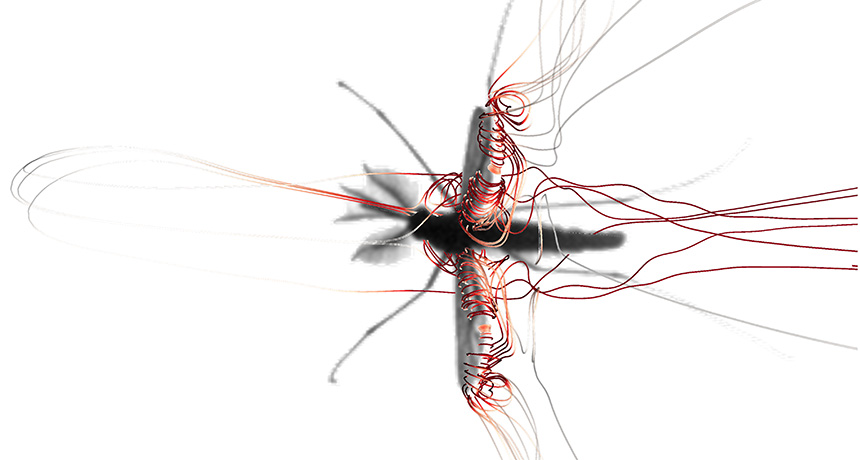
Mosquitoes take weird insect flight to new heights.
The buzzing bloodsuckers flap their long wings in narrow strokes really, really fast — more than 800 times per second in males. That’s four times faster than similarly sized insects. “The incredibly high wingbeat frequency of mosquitoes is simply mind-boggling,” says David Lentink, who studies flight at Stanford University.
Mosquitoes mostly hover. Still, it takes a lot of oomph and some unorthodox techniques to fly that slowly. Mosquitoes manage to stay aloft thanks primarily to two novel ways to generate lift when they rotate their wings, Richard Bomphrey and colleagues write March 29 in Nature. The insects essentially recycle the energy from the wake of a preceding wing stroke and then tightly rotate their wings to remain in flight. Most insects (and some birds and bats) rely on long wing strokes that create tiny low-pressure tornadoes called leading-edge vortices. The sharp front edge of the wing splits airflow in two, creating a bubble of swirling air along the front of the wing. Having low-pressure air above a wing and high-pressure air below generates lift.
But mosquitoes rapidly flap their wings up and down around a roughly 40-degree angle on average. Such short, speedy wingbeats make it impossible to generate enough lift from leading-edge vortices to stay in the air. “We knew something funny had to be going on. We just didn’t know what,” says Bomphrey, a biomechanist at the Royal Veterinary College of the University of London. So his team aimed eight high-speed cameras at hovering house mosquitoes (Culex quinquefasciatus) to model the physics of mosquito flight. It turns out the insects flap their wings in a tight figure eight formation. Leading-edge vortices generate some lift as the wings briefly cut through the air horizontally. Then, as the wings start to rotate into the curve of the figure eight, they trap the wake of the previous stroke to create another series of low-pressure swirling vortices, this time along the back edge of the wing. “This doesn’t require any power. It’s a particularly economical way of generating lift,” says Bomphrey. As the wings rotate, they also push air down, redirecting low-pressure air across the top of the wings. The wings rotate around an axis at their front edge, but if they go too far past vertical, they start to lose lift. So, the mosquito subtly shifts its wings’ turning axis from the front to the back of the wing, creating a more horizontal surface that allows the wings to continue to push air down. This also sets up the insect to benefit from the vortices along the trailing edge of the wing coming out of the turn.
Switching the axis mid-rotation “is impressive, especially since mosquito nerve cells fire just once for every few wingbeats,” says Itai Cohen, a Cornell University physicist not affiliated with the work. “Somehow this animal has evolved a complex wing stroke that takes advantage of aerodynamic forces and the mechanical infrastructure of the wing to generate complex motions with very few signals from the brain,” he says.
Bomphrey suspects that using these lift-driving forces may be common in mosquitoes and other insects that hover. But Lentink, who was not affiliated with the work, thinks it’s unlikely that lots of insects fly this way “because it seems so inefficient.”
Another force of nature may have driven mosquitoes to such illogical flight patterns: sex. Mosquito wingbeats make high-pitched tones, and males and females harmonize these tones in their search for a mate (SN: 01/31/09, p. 10). A flight style that entails fast flapping may have evolved as a result of sexual pressure to reach higher frequencies. That’s one theory anyway, and Cohen thinks it’s an interesting idea: “You’re talking about an insect sacrificing its flying capabilities in order to mate.”
It is the dazzling star of the biotech world: a powerful new tool that can deftly and precisely alter the structure of DNA. It promises cures for diseases, sturdier crops, malaria-resistant mosquitoes and more. Frenzy over the technique — known as CRISPR/Cas9 — is in full swing. Every week, new CRISPR findings are unfurled in scientific journals. In the courts, universities fight over patents. The media report on the breakthroughs as well as the ethics of this game changer almost daily.
But there is a less sequins-and-glitter side to CRISPR that’s just as alluring to anyone thirsty to understand the natural world. The biology behind CRISPR technology comes from a battle that has been raging for eons, out of sight and yet all around us (and on us, and in us).
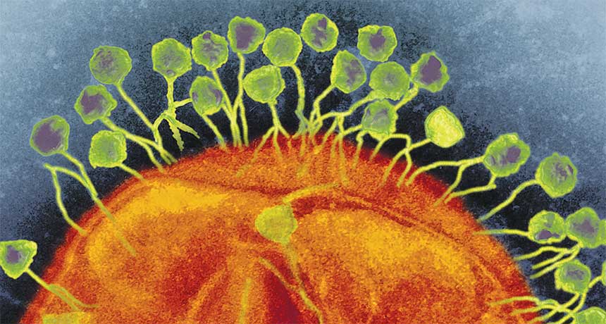
The CRISPR editing tool has its origins in microbes — bacteria and archaea that live in obscene numbers everywhere from undersea vents to the snot in the human nose. For billions of years, these single-celled organisms have been at odds with the viruses — known as phages — that attack them, invaders so plentiful that a single drop of seawater can hold 10 million. And natural CRISPR systems (there are many) play a big part in this tussle. They act as gatekeepers, essentially cataloging viruses that get into cells. If a virus shows up again, the cell — and its offspring — can recognize and destroy it. Studying this system will teach biologists much about ecology, disease and the overall workings of life on Earth.
But moving from the simple, textbook story into real life is messy. In the few years since the defensive function of CRISPR systems was first appreciated, microbiologists have busied themselves collecting samples, conducting experiments and crunching reams of DNA data to try to understand what the systems do. From that has come much elegant physiology, a mass of complexity, surprises aplenty — and more than a little mystery. Spoiled yogurt The biology is complicated, and its basic nuts and bolts took some figuring out. There are two parts to CRISPR/Cas systems: the CRISPR bit and the Cas bit. The CRISPR bit — or “clustered regularly interspaced short palindromic repeats” — was stumbled on in the late 1980s and 1990s. Scientists then slowly pieced the story together by studying microbes that thrive in animals’ guts and in salt marshes, that cause the plague and that are used to make delicious yogurt and cheese.
None of the scientists knew what they were dealing with at first. They saw stretches of DNA with a characteristic pattern: short lengths of repeated sequence separated by other DNA sequences now known as spacers. Each spacer was unique. Because the roster of spacers could differ from one cell to the next in a given microbe species, an early realization was that these differences could be useful for forensic “typing” — investigators could tell whether food poisoning cases were linked, or if someone had stolen a company’s yogurt starter culture. But curious findings piled up. Some of those spacers, it turned out, matched the DNA of phages. In a flurry of reports in 2005, scientists showed, to name one example, that strains of the lactic acid bacterium Streptococcus thermophilus contained spacers that matched genetic material of phages known to infect Streptococcus. And the more spacers a strain had, the more resistant it was to attack by phages.
This began to look a lot like learned or adaptive immunity, akin to our own antibody system: After exposure to a specific threat, your immune system remembers and you are thereafter resistant to that threat. In a classic experiment published in Science in 2007, researchers at the food company Danisco showed it was so. They could see new spacers added when a phage infected a culture of S. thermophilus. Afterward, the bacterium was immune to the phage. They could artificially engineer a phage spacer into the CRISPR DNA and see resistance emerge; when they took the spacer away, immunity was lost.
This was handy intel for an industry that could find whole vats of yogurt-making bacteria wiped out by phage infestations. It was an exciting time scientifically and commercially, says Rodolphe Barrangou of North Carolina State University in Raleigh, who did a lot of the Danisco work. “It was not just discovering a cool system, but also uncovering a powerful phage-resistance technology for the dairy industry,” he says.
The second part of the CRISPR/Cas system is the Cas bit: a set of genes located near the cluster of CRISPR spacers. The DNA sequences of these genes strongly suggested that they carried instructions for proteins that interact with DNA or RNA in some fashion — sticking to it, cutting it, copying it, unraveling it. When researchers inactivated one Cas gene or another, they saw immunity falter. Clearly, the two bits of the system — CRISPR and Cas — were a team. It took many more experiments to get to today’s basic model of how CRISPR/Cas systems fight phages — and not just phages. Other types of foreign DNA can get into microbes, including circular rings called plasmids that shuttle from cell to cell and DNA pieces called transposable elements, which jump around within genomes. CRISPRs can fend off these intruders, as well as keep a microbe’s genome in tidy order.
The process works like this: A virus injects its genetic material into the cell. Sensing this danger, the cell selects a little strip of that genetic material and adds it to the spacers in the CRISPR cluster. This step, known as immunization or adaptation, creates a list of encounters a cell has had with viruses, plasmids or other foreign bits of DNA over time — neatly lined up in reverse chronological order, newest to oldest.
Older spacers eventually get shed, but a CRISPR cluster can grow to be long — the record holder to date is 587 spacers in Haliangium ochraceum, a salt-loving microbe isolated from a piece of seaweed. “It’s like looking at the last 600 shots you had in your arm,” says Barrangou. “Think about that.”
New spacer in place, the microbe is now immunized. Later comes targeting. If that same phage enters the cell again, it’s recognized. The cell has made RNA copies of the relevant spacer, which bind to the matching spot on the genome of the invading phage. That “guide RNA” leads Cas proteins to target and snip the phage DNA, defanging the intruder. Researchers now know there are a confetti-storm of different CRISPR systems, and the list continues to grow. Some are simple — such as the CRISPR/Cas9 system that’s been adapted for gene editing in more complex creatures (SN: 4/15/17, p. 16) — and some are elaborate, with many protein workhorses deployed to get the job done.
Those who are sleuthing the evolution of CRISPR systems are deciphering a complex story. The part of the CRISPR toolbox involved in immunity (adding spacers after phages inject their genetic material) seems to have originated from a specific type of transposable element called a casposon. But the part responsible for targeting has multiple origins — in some cases, it’s another type of transposable element. In others, it’s a mystery.
The downsides Given the power of CRISPR systems to ward off foes, one might think every respectable microbe out there in the soils, vents, lakes, guts and nostrils of this planet would have one. Not so.
Numbers are far from certain, partly because science hasn’t come close to identifying all the world’s microbes, let alone probe them all for CRISPRs. But the scads of microbial genetic data accrued so far throw up interesting trends.
Tallies suggest that CRISPR systems are far more prevalent in known archaea than in known bacteria — such systems exist in roughly 90 percent of archaea and about 35 percent of bacteria, says Eugene Koonin, a computational evolutionary biologist at the National Institutes of Health in Bethesda, Md. Archaea and bacteria, though both small and single-celled, are on opposite sides of the tree of life.
Perhaps more significantly, Koonin says, almost all the known microbes that live in superhot environments have CRISPRs. His group’s math models suggest that CRISPR systems are most useful when microbes encounter a big enough variety of viruses to make adaptive memory worth having. But if there’s too much variety, and viruses are changing very fast, CRISPRs don’t really help — because you’d never see the same virus again. The superhot ecosystems, he says, seem to have a stable amount of phage diversity that’s not too high or low.
And CRISPR systems have downsides. Just as people can develop autoimmune reactions against their own bodies, bacteria and archaea can accidentally make CRISPR spacers from bits of their own DNA — and risk chewing up their own genetic material. Researchers have seen this happen. “No immunity comes without a cost,” says Rotem Sorek, a microbial genomicist at the Weizmann Institute of Science in Rehovot, Israel.
But mistakes are rare, and Sorek and his colleagues recently figured out why in the microbe they study. The researchers reported in Nature in 2015 that CRISPR spacers are created from linear bits of DNA — and phage DNA is linear when it enters cells. The bacterial chromosome is protected because of its circular form. Should it break and become linear for a spell, such as when it’s being replicated, it contains signals that ward off the Cas proteins.
There are other negatives to CRISPR systems. It’s not always a bonus to keep out phages and other invaders, which can sometimes bring in useful things. Escherichia coli O157:H7, of food poisoning fame, can make humans sick because of toxin genes it harbors that were brought in by a phage, to name just one of myriad examples. Even CRISPR systems themselves are spread around the microbial kingdom via phages, plasmids or transposable elements.
For microbes that lack CRISPR systems, there are many other ways to repel foreign DNA — as much as 10 percent of a microbial genome may be devoted to hawkish warfare, and new defense systems are still being uncovered.
Countermeasures The war between bacteria and phages is two-sided, of course. Just as a microbe wants to keep doors shut to protect its genetic integrity and escape destruction, the phage wants in.
And so the phage fights back against CRISPRs. It genetically morphs into forms that CRISPRs no longer recognize. Or it designs bespoke artillery. Microbiologist Joe Bondy-Denomy, now at the University of California, San Francisco, happened upon such customized weapons as a grad student in the lab of molecular microbiologist Alan Davidson at the University of Toronto. The team knew that the bacterium Pseudomonas aeruginosa, which lives in soil and water and can cause dangerous infections, has a vigorous CRISPR system. Yet some phages didn’t seem fazed by it.
That’s because those phages have small proteins that will bind to and interfere with this or that part of the CRISPR machinery, such as the Cas enzyme that cuts phage DNA. The binding disables the CRISPR system, the researchers reported in 2015 in Nature. Bondy-Denomy and others have since found anti-CRISPR genes in other phages and other kinds of interloping DNA. The genes are so common, Davidson says, that he wonders how many CRISPR systems are truly active.
In an especially bizarre twist, microbiologist Kimberley Seed of the University of California, Berkeley found a phage that carries its own CRISPR system and uses it to fight back against the cholera bacterium it invades, she and colleagues reported in 2013 in Nature. It chops up a segment of bacterial DNA that normally inhibits phage infection.
Of course, in this never-ending scuffle one would expect the microbes to again fight back against the phages. “It’s something I often get asked: ‘Great, the anti-CRISPRs are there, so where are the anti-anti-CRISPRs?’ ” Bondy-Denomy says. Nobody has found such things yet.
Evolution drivers It’s one thing to study CRISPR systems in well-controlled lab settings, or in just one type of microbe. It’s another to understand what all the various CRISPRs do to shape the ecosystem of a bubbling hot spring, human gut, diseased lung or cholera-tainted river. Estimates of CRISPR abundance could drop as more sampling is done, especially of dark horse microbes that researchers know little about.
In a 2016 report in Nature Communications, for example, geomicrobiologist Jill Banfield of UC Berkeley and colleagues detected 1,724 microbes in Colorado groundwater that had been treated to boost the abundance of types that are difficult to isolate. CRISPR systems were much rarer in this sample than in databases of better-known microbes.
Tallying CRISPRs is just the start, of course. Microbial communities — including those inside our own guts, where there are plenty of CRISPR systems and phages — are dynamic, not frozen. How do CRISPRs shape the evolution of phages and microbes in the wild? Banfield’s and Barrangou’s labs teamed up to watch as S. thermophilus and phages incubated together in a milk medium for hundreds of days. The team saw bacterial numbers fall as phages invaded; then bacteria acquired spacers against the phage and rallied — and phage numbers fell downward in turn. Then new phage populations sprang up, immune to S. thermophilus defenses because of genetic changes. In this way, the researchers reported in 2016 in mBio, CRISPRs are “one of the fundamental drivers of phage evolution.”
CRISPR systems can be picked up, dropped, then picked up again by bacteria and archaea over time, perhaps as conditions and needs change. The bacterium Vibrio cholerae is an example of this dynamism, as Seed and colleagues reported in 2015 in the Journal of Bacteriology. The older, classical strains of this medical blight harbored CRISPRs, but these strains went largely extinct in the wild in the 1960s. Strains that cause cholera today do not have CRISPRs.
Nobody knows why, Seed says. But scientists stress that it is a mischaracterization to paint the relationship between microbes and phages, plasmids and transposable elements as a simplistic war. Phages don’t always wreak havoc; they can slip their genomes quietly into the bacterial chromosome and coexist benignly, getting copied along with the host DNA. Phages, plasmids and transposable elements can confer new, useful traits — sometimes even essential ones. Indeed, such movement of DNA across species and strains is at the heart of how bacteria and archaea evolve.
So it’s about finding balance. “If you incorporate too much foreign DNA, you cannot maintain a species,” says Luciano Marraffini, a molecular microbiologist at the Rockefeller University in New York City whose work first showed that DNA-cutting was key to CRISPR systems. But you do need to let some DNA in, and it’s likely that some CRISPR systems permit this: The system he studies in Staphylococcus epidermidis, for example, only goes after phages that are in their cell-killing, or lytic, state, he and colleagues reported in 2014 in Nature. Beyond defense One thing is very clear about CRISPR systems: They are perplexing in many ways. For a start, the spacers in a microbe should reflect its own, individual story of the phages it has encountered. So you’d think there would be local pedigrees, that a bacterium sampled in France would have a different spacer cluster from a bacterium sampled in Argentina. This is not what researchers always see.
Take the nasty P. aeruginosa. Rachel Whitaker, a microbial population biologist at the University of Illinois at Urbana-Champaign, studies Pseudomonas samples collected from people with cystic fibrosis, whose lungs develop chronic infections. She’s found no sign that two patients living close to each other carry more-similar P. aeruginosa CRISPRs than two patients thousands of miles apart. Yet surely one would expect nearby CRISPRs to be closer matches, because the Pseudomonas would have encountered similar phages. “It’s very weird,” Whitaker says.
Others have seen the same thing in heat-loving bacteria sampled from very distant bubbling hot springs. It’s as if scientists don’t truly understand how bacteria spread around the world — there could be a strong effect of far-flung passage by air or wind, says Konstantin Severinov, who studies CRISPR systems at Rutgers University in New Brunswick, N.J. Another weirdness is the differing vigor of CRISPR systems. Some are very active. Molecular biologist Devaki Bhaya of the Carnegie Institution for Science’s plant biology department at Stanford University sees clear signs that spacers are frequently added and dropped in the cyanobacteria of Yellowstone’s hot springs, for example. But other systems are sluggish, and E. coli, that classic workhorse of genetics research, has a respectable-looking CRISPR system — that is switched off.
It may have been off for a long time. Some 42,000 years ago, a baby woolly mammoth died in what is now northwestern Siberia. The remains, found in 2007, were so well-preserved that the intestines were intact and E. coli DNA could be extracted.
In research published in Molecular Ecology in January, Severinov’s team found surprising similarities between the spacers in the mammoth-derived E. coli CRISPR cluster and those in modern-day E. coli. “There was no turnover in all that time,” Severinov marvels. If the CRISPR system isn’t active, why does E. coli bother to keep it?
That quandary leads neatly to what some researchers refer to as an intellectually “scandalous situation.”
In some cases, the genetic sequence of spacers nicely matches phage DNA. But overall, only a fraction (around 1 to 2 percent) of the spacers scientists know about have been matched to a virus or a plasmid. In E. coli, the spacers don’t match common, classic phages known to infect the bacterium. “Is it the case that there is a huge, unknown amount of viral dark matter in the world?” says Koonin — or are phages evolving superfast? “Or is it something completely different?”
Faced with this conundrum, some researchers strongly suspect — and have evidence — that CRISPR systems may do more than defend; they may have other jobs. Communication, perhaps. Or turning genes on and off.
But some microbes’ CRISPR sequences do make sense, especially if looking at the spacers most recently added, and others may be clues to phages still undiscovered. So even as they scratch their heads about many things CRISPR, scientists are also excited by the stories CRISPR clusters can tell about the viruses and other bits of DNA that bacteria and archaea encounter and that they choose, for whatever reason, to note for the record. What do microbes pay attention to? What do they ignore?
CRISPRs offer a bright new window on such questions and, indeed, already are unearthing novel phages and facts about who infects whom in the microscopic world.
“We can catalog everything that’s out there. But we don’t really know what matters,” says Bondy-Denomy. “CRISPRs can help us understand.”
A severe coral bleaching event spurred by high ocean temperatures has struck the Great Barrier Reef for an unprecedented second time in 12 months, reveal aerial surveys released April 10 by scientists at James Cook University in Townsville, Australia. While last year the northern third of the reef was hardest hit, this time around the reef’s midsection experienced the worst bleaching. The two bleaching events together span around 1,500 kilometers of the 2,300-kilometer-long reef.
“It takes at least a decade for a full recovery of even the fastest growing corals, so mass bleaching events 12 months apart offers zero prospect of recovery for reefs that were damaged in 2016,” James Kerry, one of the researchers behind the finding, said in a statement.
Bleached corals aren’t dead. Trauma, disease or warm water can cause an exodus of the symbiotic algae that provide corals with food and vibrant color schemes. If better conditions, such as cooler waters, return, the algae may return to their homes. If they don’t come back, though, the corals starve.
Warming caused by El Niño exacerbated last year’s bleaching event. With El Niño now long gone, the researchers blame this year’s bleaching largely on global warming. If humans don’t curb emissions of planet-warming greenhouse gases, scientists warn that the entire reef could be in jeopardy.
A handful of measurements of decaying particles has seemed slightly off-kilter for years, intriguing physicists. Now a new decay measurement at the Large Hadron Collider in Geneva has amplified that interest into tentative enthusiasm, with theoretical physicists proposing that weird new particles could explain the results. Scientists with the LHCb experiment reported the new result on April 18 in a seminar at the European particle physics lab CERN, which hosts the LHC.
“It’s incredibly exciting,” says theoretical physicist Benjamin Grinstein of the University of California, San Diego. The new measurement is “a further hint that there’s something new and unexpected happening in very fundamental interactions.” Other physicists, however, are more cautious, betting that the series of hints will not lead to a new discovery. “One should always remain suspicious of an effect that does not show up in a clear way” in any individual measurement, Carlos Wagner of the University of Chicago wrote in an e-mail.
Taken in isolation, none of the measurements rise beyond the level that can be explained by a statistical fluctuation, meaning that the discrepancies could easily disappear with more data. But, says theoretical physicist David London of the University of Montreal, there are multiple independent hints, “and they all seem to be pointing at something.”
The measurements all involve a class of particle called a B meson, which can be produced when protons are smashed together in the LHC. When a B meson decays, it can produce a type of particle called a kaon that is accompanied either by an electron and a positron (an antimatter version of an electron) or by a muon — the electron’s heavier cousin — and an antimuon.
According to physicists’ accepted theories, muons and electrons should behave essentially identically aside from the effects of their differing masses. That means the two kinds of particles should have an even chance of being produced in such B meson decays. But in the new result, the scientists found only about seven decays with muons for every 10 with electrons.
There are several varieties of B mesons. All are made up of one quark — a type of fundamental particle that also makes up protons and neutrons — and one antiquark. One of the two particles is a type called a “bottom” quark (or antiquark), hence the B meson’s name. Earlier measurements of another variety of B meson decay also found a muon shortage. What’s more, measurements of the angles at which particles are emitted in some types of B meson decay also appear slightly out of whack, adding to the sense that something funny may be going on in such decays.
“We are excited by how [the measurements] all seem to fit together,” says LHCb spokesperson Guy Wilkinson, an experimental physicist at the University of Oxford in England. If more data confirm that B mesons misbehave, a likely explanation would be a new particle that interacts differently with muons than it does with electrons. One such particle could be a leptoquark — a particle that acts as a bridge between quarks and leptons, the class of particle that includes electrons and muons. Or it could be a heavy, electrically neutral particle called a Z-prime boson.
Physicists spawned a similar hubbub in 2016, when the ATLAS and CMS experiments at the LHC saw hints of a potential new particle that decayed to two photons (SN: 5/28/16, p. 11). Those hints evaporated with more data, and the current anomalies could do likewise. Although the two sets of measurements are very different, says Wolfgang Altmannshofer of the University of Cincinnati, “from the point of the overall excitement, I would say the two things are roughly comparable.”
Luckily, LHCb scientists still have a lot more data to dig into. The researchers used particle collisions only from before 2013, when the LHC was running at lower energy than it is now. “We have to get back to the grindstone and try and analyze more of the data we have,” says Wilkinson. Updated results could be ready in about half a year, he says, and should allow for a more definitive conclusion.